Condensed Matter & Complex Systems
Find out more about PhD projects in Condensed Matter & Complex Systems research via the research themes below.
Astrobiology
We investigate life in extreme environments, how life adapts to single and multiple extremes, how life adapts to conditions in the planetary crust and we investigate the habitability of other planetary bodies. Our work involved field, laboratory and theoretical approaches. Please contact us about current opportunities.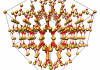
Computational Materials Physics
We use computer simulations to study the properties of materials. By applying methods ranging from electronic structure calculations to classical molecular dynamics, we study liquids, colloids, and atomic or molecular crystals under different temperature and pressure conditions, both in and away from equilibrium.
Extreme Conditions Physics
We study how materials react and change when being subjected to extremes of pressure, temperature, fields and strain. We use diamond anvils or large lasers to create high pressures, liquid He cooling and laser heating to change temperatures, and x-rays, neutrons and optical spectroscopy to study how the materials change. Understanding our observations is aided by computation and simulation, using methods such as electronic structure calculations, classical and quantum molecular dynamics.
Physics of Living Matter
We use experiments, computer simulations, and theoretical calculations to understand how physical laws affect living organisms.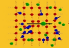
Quantum Ordering
Quantum Order refers to correlated states of matter that have a quantum origin, including different forms of superconductivity, magnetism, ferroelectricity, and topologically protected states. Many of these states emerge at low temperatures where quantum effects become more pronounced. Many also require pure single crystals. We grow our own crystals, which allows us to lead the hunt for new forms of order, which we study down to very low temperature and in high magnetic fields in-house and at central facilities with neutrons, X-rays and muons.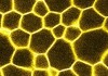
Soft Matter Physics
"Soft matter" is a convenient term for materials that are easily deformed by thermal fluctuations and external forces. In short, it refers to ‘all things squishy’! Everyday examples include paint, blood, milk, spreads and ice cream. Soft materials share several characteristic features, e.g. that their building blocks are intermediate in size between atoms and grains, and this is crucial to understanding their behaviour. In the Edinburgh Soft Matter Physics group, we use experiments, computer simulations, and theoretical calculations to understand colloidal and granular model systems for phenomena ranging from jamming to bacterial colonies and to rationally design novel soft materials for use in applications ranging from foods to energy materials.